
fluorescence microscopy
- noun
- /flɔːˈrɛsəns maɪˈkrɒskəpi/
- Specialized
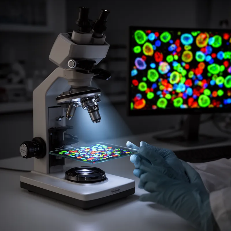
- Photo (color): Using fluorescence microscopy, chemist Edward Lulai determines cellular changes during wound healing in a damaged potato.
Examples
-
Using fluorescence microscopy, the team was able to observe how the cells reacted to the treatment.
-
By 1982, we established a method for detecting small DNA molecules (approximately 1 kb) in organelles in vivo using high-resolution fluorescence microscopy.
Academic text (1996) -
The scientists applied fluorescence microscopy to examine the intricate details of the sample's cellular structure.
-
The research relied on fluorescence microscopy to visualize the fluorescent markers in the tissue samples.
Surface Forms
Morphology
The phrase is transparently compositional: it denotes a microscopy technique that uses fluorescence (light emitted after excitation) to visualize specimens. Because both constituents directly describe the method, a B1 learner who knows the words 'fluorescence' and 'microscopy' can reliably infer the MWE's meaning; this is a common noun–noun technical compound found across languages.
Etymology
The term fluorescence microscopy comes from fluorescence, which means to 'glow' when hit by special light, and microscopy, which means looking at very small things with a microscope. Scientists shine that special light to make dyes or parts of cells glow and then use a microscope to see them, so the phrase means 'using glowing dyes and a microscope to view tiny structures'.

